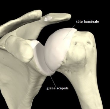
Bone parts articulated between them are covered with cartilage, soft and slippery tissue ‘oiled’ by the synovial fluid. The boundaries are constituted by a membrane called joint capsule and fibrous sustaining structures called ligaments.
On a frontal radiograph, the two bones seem separated by a space (= joint space), in fact it is the thickness of the cartilage which is radiolucent (invisible on X-ray). The disappearance of this space is called ‘pinching’ of the joint space corresponds to the first radiological sign of osteoarthritis.
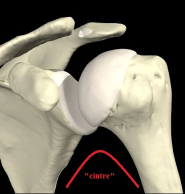
The scapulo-humeral (or omo-humeral) hanger is described by the fact that the lower curvature of the scapula (scapula) is harmoniously and almost symmetrically extended by the neck of the humerus.
This hanger is present in the normal state, when it is broken it corresponds to an upper migration of the humeral head by disappearance of the rotator cuff.
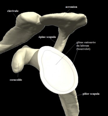
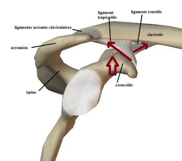
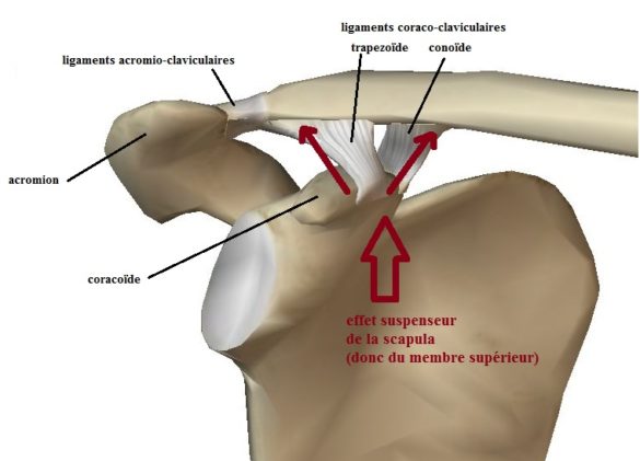
The Acromioclavicular joint corresponds to the bony ceiling of the shoulder, at the level of the external termination of the clavicle in relation to the acromion which terminates the spine of the scapula. This joint has an articular capsule and clean ligaments ‘acromio-clavicular’ which provides stability especially anteroposterior, but the particularity of this joint lies in the fact that it is stabilized in the vertical direction by two bulky large ligaments ‘coraco-clavicular’ which unite the lower surface of the external third of the clavicle to the coracoid, V-shaped.
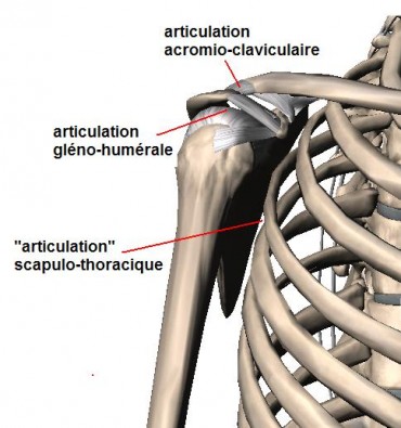
The acromioclavicular joint is a secondary shoulder joint compared to the glenohumeral.
It combines the acromion, the terminal bone appendix of the scapula spine (bone ceiling of the shoulder and area of insertion of the deltoid muscle) and the lateral end of the clavicle (upper thorax bumper).
This joint does not participate directly in the movements of the shoulder but it is extremely stressed during the use of the member, especially during movements at or beyond the plane of the shoulder.
As in any joint there are articular surfaces covered with cartilage, but it also includes a damping element of stress-related constraints called ‘pseudo-meniscus’
(Like the knee meniscus absorbs the constraints of the ground support).
It also has ligaments that stabilizes it:
- acromioclavicular ligaments: direct stabilizers of the joint, related to the articular capsule.
- coraco-clavicular ligaments: not directly related to the joint, in fact stabilizers of the clavicle-scapula pair (via the acromion). These ligaments are called ‘suspensors’ of the scapula and also of the upper limb, because in their absence there is a fall of the stump of the shoulder caused by the weight of the limb, realizing the acromioclavicular dysjunction (and not the ascension). clavicle).
Two types of pathology are common in this joint:
- degenerative or inflammatory pathology: related to joint overuse, inflammatory joint disease or distant post-traumatic disease;
- direct traumatic pathology: sprains and acromioclavicular dysjunctions.
Sterno-clavicular joint
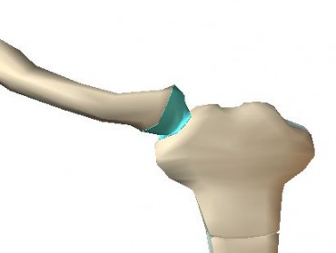
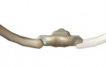
The medial (internal) end of the clavicle articulates with the sternum but also the first rib (its origin at this level is cartilage).
This joint includes an articular capsule, own ligaments (sterno-clavicular) and an articular disc (pseudo-meniscus) as well as the acromioclavicular joint.
It constitutes a zone of damping of the stresses transmitted by the clavicle to the thorax (by the sternum) and coming from the upper limb.
The relations with the big vessels of the base of the neck are narrow (venous trunks in particular).
Normal anatomy positions on the same clavicle and sternum axis.
Sterno-clavicular pathologies are dominated by:
- Traumatology: Sternoclavicular sprains and luxations (including retro-sternal luxation)
- degenerative pathology often by joint overwork or rheumatoid arthritis
These pathologies are briefly explained in a chapter dedicated to pathologies-> other pathologies-> sternoclavicular
Scapulo-thoracic articulation is not in the strict sense of the term an articulation.
It actually represents a sliding space between the thoracic wall (the costal grill) and the anterior surface of the scapula.
A serous bursa allows this sliding.
The scapulo-thoracic mobilities are related to the sliding and rocking of the body of the scapula on thorax.
The scapula remains ‘plated’ on the thorax via the anterior serrated muscle (innervated by the long thoracic nerve).
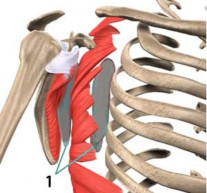
We use the term articulation because the movements in question are far from being innocuous!
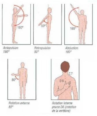
The total mobility of the shoulder (arms vertically, rotations) corresponds to an associated mobility of the glenohumeral and scapulothoracic.
There are also lateral and superior movements of the scapula as shown in the diagram opposite.
To be convinced of this it is necessary to block the movements of the scapula by holding it firmly by a hand on the top of the shoulder, then the shoulder does not go up to the vertical …
The arm above the shoulder implies a rocking of nearly 180 ° of the scapula.
diseases
The pathologies of scapulothoracic are less frequent and especially less clearly identified than other joints.

