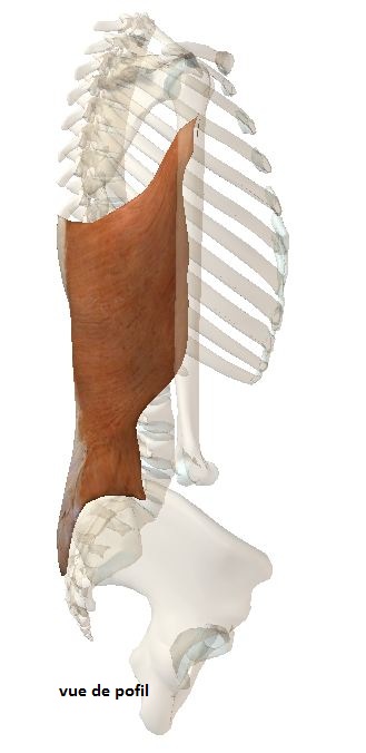
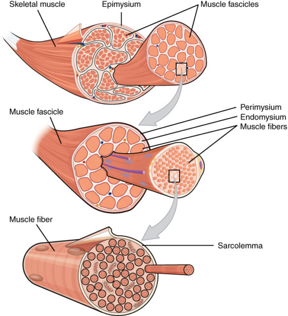
It is a very organized and powerful structure to create a movement of one piece of bone on another through a joint between the two bones. We will only be interested in skeletal striated muscles in these chapters (because they alone allow voluntary movement via a motor nerve), we will not talk about the smooth muscles that are found elsewhere in the human body whose functioning is not voluntary, nor the heart muscle which is also striated but of a different mode of operation …)
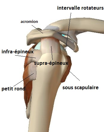
In addition, the edges of the sub-scapular and supraspinous tendons delineate a passage opening of the tendon of the long portion of the biceps called the bicipital groove situated in a depression limited by the bony tuberosities of the humerus.
There are 4 muscles, 4 tendons:
the subscapularis muscle forward
supra-thorny muscle (supraspinous) above
intraspinous (infraspinous) muscles and small round back
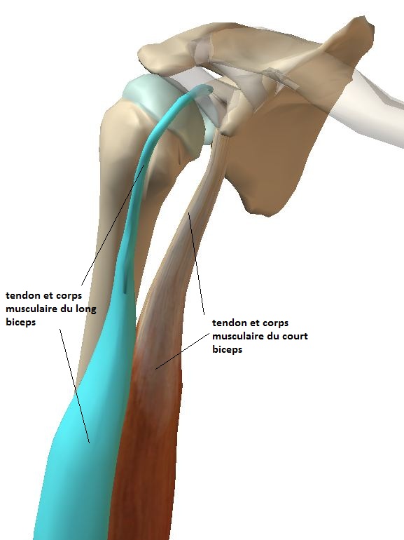
The brachial biceps is the superficial anterior muscle of the arm playing a role in the shoulder.
International Common Name: ‘Biceps Brachii’ (in Latin)
It is constituted by 2 muscular bundles (‘bi’ ceps).
Short portion
The short portion (short Biceps), the innermost, which starts from the coracoid, gives a fleshy internal muscular body that is common with that of the coraco-brachialis muscle on the first centimeters (= tendonconjoint coracobiceps) then its muscular body joins the one resulting of the long portion of the biceps.
This common tendon and the corresponding muscular body are useful in stabilizing the shoulder when performing a shoulder stop.
They also have relationships with the musculocutaneous nerve
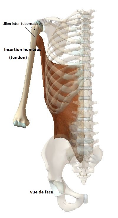
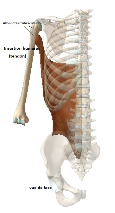
The long dorsal is a powerful muscle of the trunk and shoulder among the largest of the human body, muscle ‘climb’.
Called ‘Latissimus dorsi’ in Latin in international naming
l intervenes in the shoulder by its tendon inserting on the medial embroidery (internal) of the interveberular groove (intimate with the large round tendon).
Its origin is dorsal multiple and dispersed: from the sacrum, the iliac wing (pelvis), the spines of the vertebrae and some ribs passing (inconstantly) by the lower corner of the scapula.
In addition to climbing, it participates in the internal rotation of the arm (hand in the back) and adduction arm (brings the arm of the axis of the body).
This muscle has a special vasularization and dimensions such that it is very useful in plastic surgery and reconstruction (breast removal suites, coverage of dilapidated members etc ..)
It is used as a tendon transfer (replacement of a non-repairable tendon by another), especially in major ruptures of the rotator cuff.
We distinguish the pectoralis major muscle and the pectoralis minor muscle
Pectoralis Major Muscle:
pectoralis major in Latin (= international non-proprietary name)
Muscle superficiel de la région antérieur du thorax
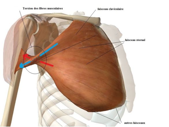
Includes several bundles (or heads) of origin:
– cluster (head) clavicular,at the internal 2/3 level of the clavicle
– sternal bundle
– chondro-costal bundle (first 6 ribs)
– Abdominal bundle (rectus muscle)
Terminaison
It ends with a J-shaped tendon on the inner edge of the bicipital groove of the humerus.
There is a twisting of the muscle fibers when they end in a short tendon.
Fonction
Adductor (places the arm medially towards the opposite arm)
Internal Rotator
anti-thruster (anterior elevation)
Innervation
By the handle of the pectorals
This muscle can be used for tendon transfers in the context of unrepairable rotator cuff tears
(usually a clavicle or sternal muscle bundle) to replace the subscapularis tendon.
Small pectoral muscle
Pectoralis minor in Latin (= international non-proprietary name)
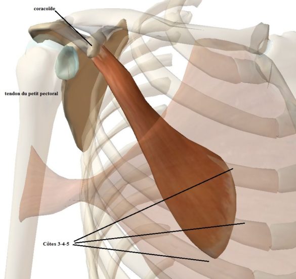
Located below the pectoralis major, flat and triangular.
Has as origin the 3rd, 4th and 5th ribs
Ends with a very short tendon that fits on the coracoid process.
Also innervated by the pectoral handle.
It is an internal rotator and an antipulsor but also an accessory muscle of respiration (respiratory insufficiency).
The tendon is sytematically divided when the coracoid is removed as part of a Shoulder Stop operation.
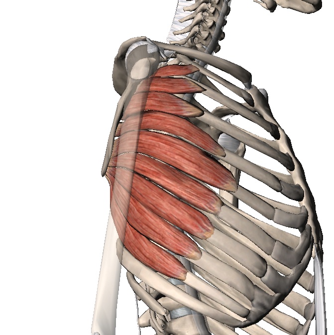
Anterior serrated
Otherwise called Serratus anterior in Latin (international nonproprietary name).
Description
Particular muscle if any, composed of multiple muscular bodies in the form of fingers = fingerings)
Origin: the first 9 ribs
End : the medial (internal) edge of the scapula (scapula)
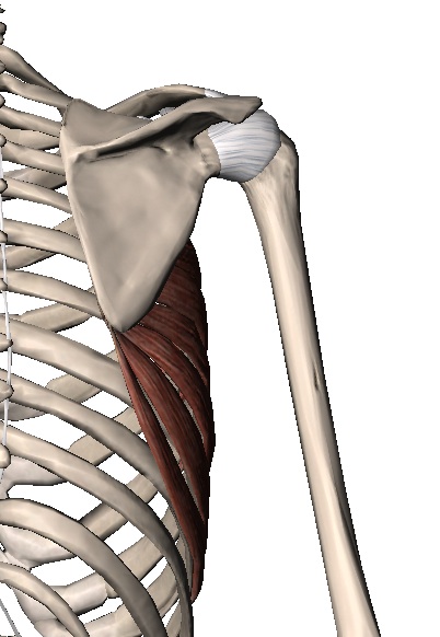
His role is to hold the scapula against the costal grill (from which it is separated by sliding spaces) during the movements of the arm forward and upwards; in the company of the other stabilizers of the scapula (Trapeze, Rhomboids, Elevator of the scapula, incidentally Small pectoral).
He is innervated by the long thoracic nerve (or Charles Bell’s nerve) that comes directly from the brachial plexus.Its paralysis causes a form of detachment of the trunk scapula called ‘sacula alata’ (winged scapula or Winging scapula in English).
In reconstructive surgery it is used to cover loss of substance especially in the limbs (bone discovered), this concerns only the last 3 fingerings.

